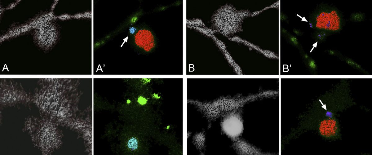
Images of Mitochondria. Four slides showing the progression/evulsion of dendritic arbors of ganglion cells. Black background. Gray dendritic structure with sporadic splotches of red and bright green.

NICKELLS LAB

Welcome to the Nickells Lab


The primary focus of the lab is to study the molecular pathology of retinal ganglion cell death after injury to the optic nerve with an ultimate goal to develop therapeutic approaches to attenuate ganglion cell death in common optic nerve diseases like glaucoma. A principal area of research is to understand the roles played by the BCL2 gene family of proteins, which control the intrinsic apoptotic pathway. Our research employs a variety of mouse models of disease including controlled acute optic nerve injury and chronic inherited glaucoma.
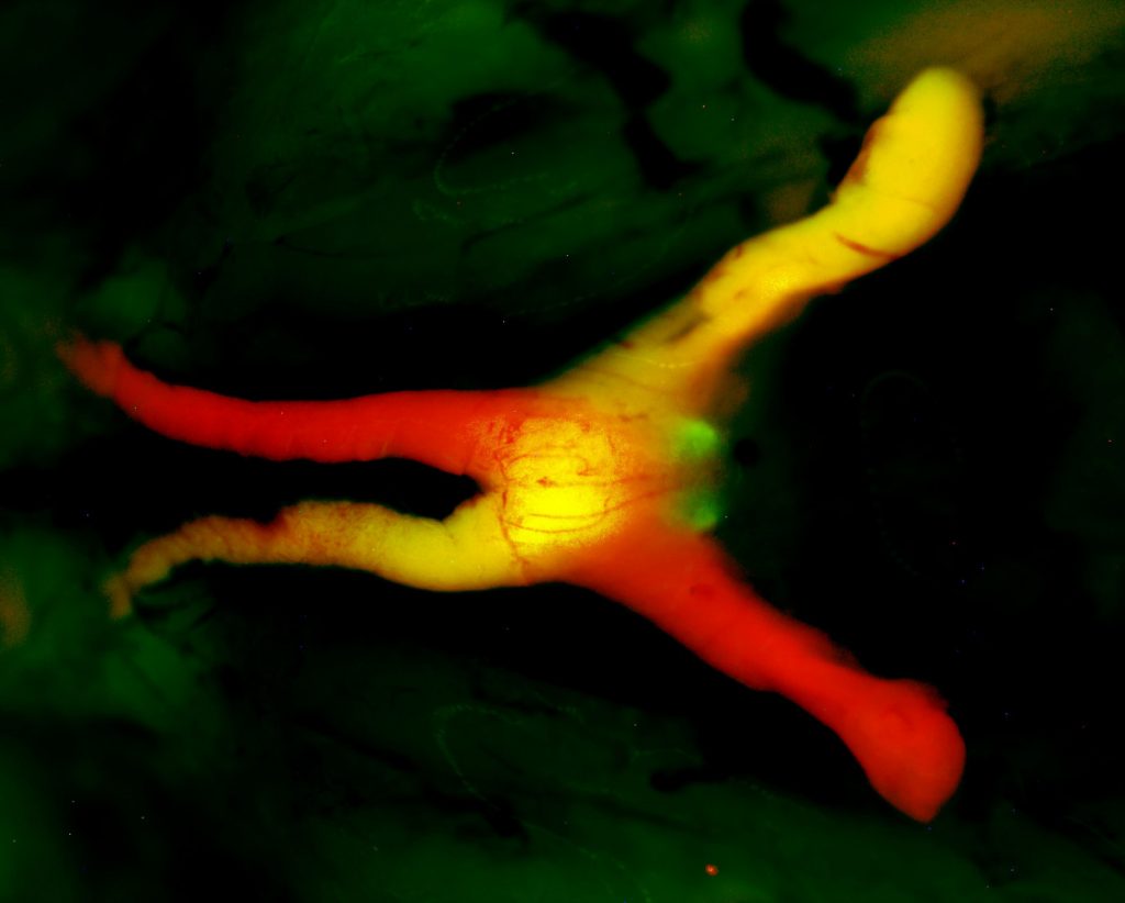
 

Glowing red and yellow nerve branching over a black and green mottled background.
This image show fluorescent labeling of the exposed optic tract in a mouse that has been treated with adeno-associated virus (serotype 2/2) to deliver a mCherry-BCLXL fusion protein (red). One of the tracts has also been labeled by previous injection of cholera toxin B protein conjugated to Alexa488 (green).