
Quiescent Retinal Ganglion Cells — The Zombie Apocalypse
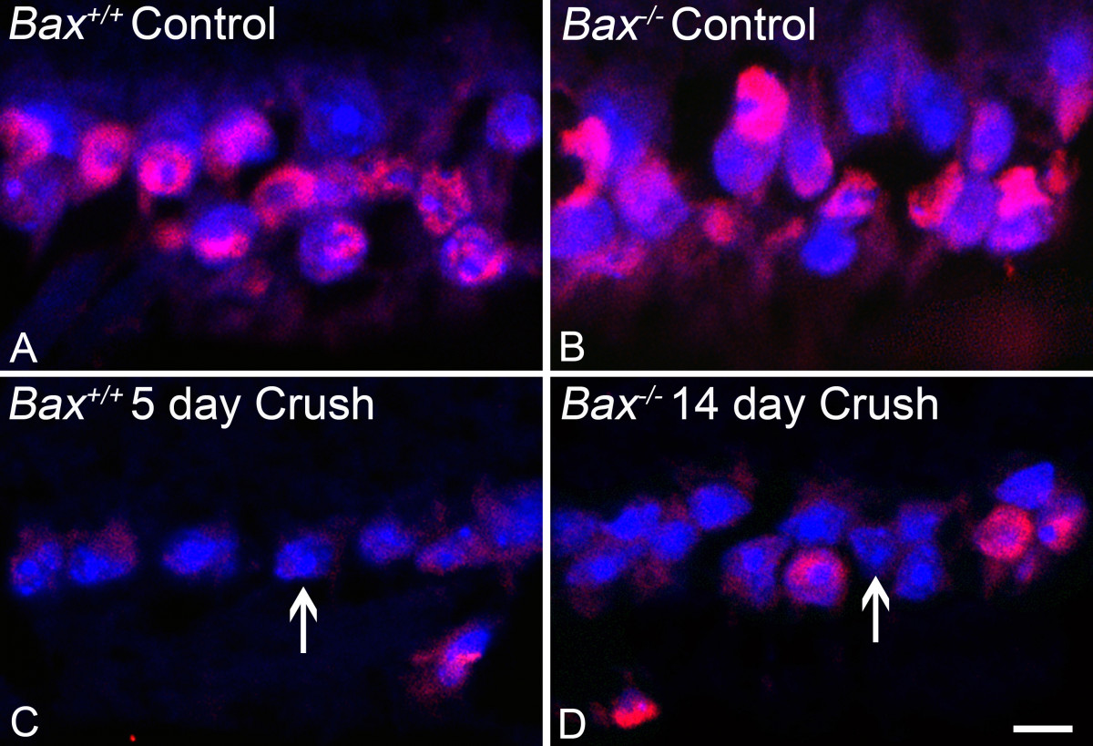
A principal goal in developing a neuroprotective strategy is to block the death of the cells and also have them retain normal function. A major hurdle in this development is the early onset of chromatin modifications that function to reset the genome of damaged retinal ganglion cells such that normal gene expression is globally silenced. As a consequence, even “protected” ganglion cells no longer express the genes they require to function normally, a condition we equate to the formation of zombie cells. Our studies have focused on the process of widespread histone deacetylation by the activation of enzymes called histone deacetylases (HDACs). HDAC activity early in apoptosis contributes to the formation of heterochromatin that helps to block active gene expression and we have found that deletion of select HDACs, or treatment with potent HDAC inhibitors, can help prevent heterochromatin formation and/or restore normal gene expression. Currently, we are exploring if HDAC inhibitors can reactivate quiescent (zombie) ganglion cells that have survived after acute optic nerve damage.
Selected Reading
- Donahue RJ, Maes ME, Nickells RW. BAX depleted retinal ganglion cells survive and become quiescent following optic nerve damage. Mol. Neurobiol. 57:1070-1084, 2020.
- Janssen KT, Mac Nair CE, Dietz JA, Schlamp CL, Nickells RW. Nuclear atrophy in dying retinal ganglion cells precedes the Bax-dependent stage of apoptosis. Invest. Ophthalmol. Vis. Sci. 54:1805-1815, 2013.
- Pelzel HR, Schlamp CL, Nickells RW. Histone H4 deacetylation plays a critical role in early gene silencing during neuronal apoptosis. BMC Neurosci. 11:62, 2010.
- Schmitt HM, Pelzel HR, Schlamp CL, Nickells RW. Histone deacetylase 3 (HDAC3) plays an important role in retinal ganglion cell death after acute optic nerve injury. Mol. Neurodegen. 9: 39, 2014.
- Schmitt HM, Fehrman RL, Maes ME, Yang H, Guo LW. Schlamp CL, Pelzel HR, Nickells RW. Increased susceptibility and intrinsic apoptotic signaling in neurons by forced HDAC3 expression. Invest. Ophthalmol. Vis. Sci. 62:14, 2021.